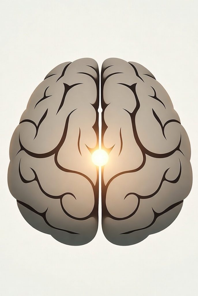
The pineal gland, a small pine cone-shaped endocrine gland in the brain’s epithalamus, produces melatonin—a serotonin-derived hormone that regulates sleep, circadian rhythms, and light-dark cycles. Located near the third ventricle and connected by the pineal stalk, it lacks a blood-brain barrier as a circumventricular organ. Pinealocytes synthesize melatonin, peaking at night under sympathetic control from the superior cervical ganglion. It develops in utero, grows until age 2, and often calcifies with age via corpora arenacea (brain sand), potentially reducing function. Historically called the “third eye” or “seat of the soul” by René Descartes, it evolved from light-sensitive parietal eyes in reptiles. Rare pineal tumors cause hydrocephalus or precocious puberty; melatonin aids sleep disorders, immunity, and antioxidant defense. In animals, it drives seasonal reproduction and photoperiod responses.
Long Version
The pineal gland, also known as the pineal body or epiphysis cerebri, is a small, unpaired endocrine gland within the brain that plays a crucial role in the endocrine system. Situated in the epithalamus, it produces melatonin, a serotonin-derived hormone that regulates sleep patterns, circadian rhythms, and responses to the light-dark cycle. Often described as pine cone-shaped, the gland has been historically linked to metaphysical concepts such as the third eye or seat of the soul, notably by philosopher René Descartes. As a circumventricular organ, it lacks a blood-brain barrier, allowing direct interaction with circulating substances. Found in most vertebrates, its evolutionary origins trace back to photoreceptive structures like the parietal eye or pineal eye in lower animals, highlighting its role in adapting to photoperiod changes.
Anatomy
Location
The pineal gland resides in the midline of the brain, posterior to the third ventricle and nestled between the two thalamic bodies. It attaches via the pineal stalk to the roof of the diencephalon, positioned in a groove where the thalamic halves meet, just above the superior colliculi and below the splenium of the corpus callosum. The gland projects into the pineal recess, a cerebrospinal fluid-filled extension of the third ventricle, and lies adjacent to the habenular commissure and quadrigeminal cistern. This central placement ensures its integration with key neural pathways, including those from the superior cervical ganglion for sympathetic innervation.
Structure
Measuring approximately 5–8 mm in length and weighing about 0.1 g in adult humans, the reddish-gray pineal gland consists of lobular parenchyma made up of pinealocytes—specialized endocrine cells with 4–6 processes and lightly basophilic cytoplasm—surrounded by connective tissue spaces and a pial capsule. Supporting interstitial cells with elongated nuclei, perivascular phagocytes for antigen presentation, and in some species, pineal neurons or peptidergic neuron-like cells contribute to its cellular composition. The gland’s high cellularity can sometimes mimic neoplasms on imaging. In adults, it often contains corpora arenacea, or brain sand, which are calcified deposits increasing with age. To enhance understanding, note that these structural elements allow the gland to function as both an endocrine producer and a sensor of environmental cues, with pinealocytes containing dense-core vesicles for hormone storage and release.
Blood Supply and Innervation
The pineal gland receives abundant blood flow, second only to the kidney among organs, via choroidal branches of the posterior cerebral artery, with drainage through the internal cerebral vein. Its permeable capillaries, characteristic of circumventricular organs, facilitate solute exchange. Innervation includes sympathetic fibers from the superior cervical ganglion via the nervus conarii, parasympathetic inputs from the pterygopalatine and otic ganglia, and trigeminal ganglion projections involving pituitary adenylate cyclase-activating peptide (PACAP). This neural pathway links retinal light detection to melatonin synthesis: intrinsically photosensitive retinal ganglion cells signal the suprachiasmatic nucleus, which relays via the paraventricular nucleus and spinal cord to the superior cervical ganglion, ultimately modulating gland activity. Enhancing this detail, the high vascularity supports rapid hormone dissemination, crucial for timely responses to diurnal changes, and recent studies emphasize how disruptions in this innervation can contribute to mood disorders linked to circadian misalignment.
Development
Embryologically, the pineal gland arises from neural ectoderm on the roof of the diencephalon during the seventh week of gestation, forming as an evagination from the caudal portion of the third ventricle roof. It matures into tubules of innervated cells separated by connective tissue, reaching full development by the first decade of life. The gland enlarges from birth to around age 2, stabilizing thereafter, with subtle asymmetry in some species like zebrafish. In humans, high childhood melatonin levels inhibit sexual development, decreasing at puberty; disruptions can lead to precocious puberty. To enhance, developmental studies reveal that genetic factors, such as mutations in melatonin pathway genes, can influence gland formation and contribute to neurodevelopmental conditions, underscoring its role beyond mere hormone production.
Function
The pineal gland’s primary role is synthesizing melatonin, a serotonin-derived hormone from tryptophan, which governs circadian rhythms and sleep-wake cycles in response to the light-dark cycle. Production peaks in darkness, inhibited by light via the retinohypothalamic tract, with the enzyme serotonin N-acetyltransferase (NAT) as the rate-limiting step. Melatonin influences the suprachiasmatic nucleus, retina, and pituitary pars tuberalis, binding to MT1 and MT2 receptors to regulate sleep, immune function, and antioxidant activity. In rodents, it modulates pituitary secretion of follicle-stimulating hormone (FSH) and luteinizing hormone (LH), affects bone metabolism, and protects against neurodegeneration. Additional secretions like pinoline (controversial) and potential neurosteroids underscore its broader endocrine impact. Recent research links melatonin to cholesterol metabolism, serum element balance (e.g., zinc), and cardioprotection, with heart diseases causing immune-mediated denervation and reduced melatonin, exacerbating sleep disturbances. Enhancing this section, emerging evidence suggests melatonin’s anti-inflammatory properties may aid in mitigating oxidative stress in conditions like diabetes and cancer, positioning the gland as a key player in systemic health regulation.
Calcification
Calcification of the pineal gland begins in childhood, with corpora arenacea (brain sand) deposits of calcium, magnesium, and ammonium phosphates accumulating by young adulthood in up to 40% of individuals. Visible on X-rays, these calcifications impair melatonin synthesis, potentially disrupting sleep and circadian rhythms, though evidence is mixed. Higher rates correlate with aging, Alzheimer disease, and geographic variations, serving as radiographic landmarks but loosely associated with migraines or insomnia. To enhance, lifestyle factors such as fluoride exposure or poor diet may accelerate calcification, and interventions like increased antioxidant intake are being explored to preserve gland function.
Clinical Significance
Disorders of the pineal gland include reduced volume in obesity, insomnia, and multiple sclerosis (notably in females), linking to melatonin deficits and immune dysregulation. Pineal tumors, such as pinealomas, pineoblastomas, pineocytomas, germinomas, or mixed types, are rare (less than 1% of brain tumors) but challenging due to location, often causing hydrocephalus from aqueduct compression, Parinaud syndrome, headaches, visual disturbances, or endocrine issues like precocious puberty. Craniopharyngiomas may involve adjacent structures. Diagnosis uses CSF/serum markers (e.g., beta-HCG, AFP) and MRI; treatment involves surgery, radiation, chemotherapy, or hydrocephalus management via ventriculostomy or shunts. Melatonin therapy aids circadian disorders like jet lag or delayed sleep phase, with genetic ASMT mutations tied to autism and ADHD. Recent research highlights tumor marker diagnostics (e.g., microRNAs) and melatonin’s role in metabolic and cardiac health. Enhancing here, advancements in minimally invasive neurosurgery and targeted melatonin analogs are improving outcomes for gland-related pathologies, with ongoing trials exploring its potential in neurodegenerative disease prevention.
History
Ancient descriptions by Herophilus (3rd century BCE) viewed the pineal gland as a valve for vital spirits, while Galen (2nd century CE) named it “konario” for its pine cone-shaped form, dismissing functional roles. René Descartes (17th century) proposed it as the seat of the soul for mind-body interaction, critiqued by Thomas Willis. 19th-century studies by Spencer and Leydig identified the parietal eye in lizards, with Holmgren noting photoreceptor qualities. Melatonin isolation by Aaron Lerner in 1958 advanced chronobiology. Historical links to mental disorders persist, with pineal extracts tested for behavioral effects in the 1930s. Bibliometric analyses show rising research since the 1970s, peaking in melatonin, circadian rhythms, and aging studies. To enhance, modern interpretations blend historical mysticism with scientific inquiry, influencing fields like neurophilosophy and bioethics.
In Other Animals
Conserved across vertebrates except hagfish and protochordates, the pineal gland evolved from paired photosensory organs, evident in fossil skulls with parietal foramina. In amphibians and reptiles, it connects to the parietal eye (pineal eye), a direct photoreceptor regulating photoperiod-dependent behaviors. Birds and rodents exhibit complex shapes, with melanopsin expression enabling light-sensitive circadian control akin to the mammalian suprachiasmatic nucleus. In polar mammals, it enlarges for seasonal adaptations; pinealectomy impairs reproduction in seasonally breeding species. Recent genetic studies in zebrafish reveal bsx protein’s role in left-right asymmetry and melatonin cycles, underscoring evolutionary ties between asymmetry and day-night regulation. Enhancing this, comparative anatomy reveals how the gland’s photosensitivity in non-mammals informs human therapies for light-related disorders, such as seasonal affective disorder.